By Gregg A. DuPont DVM FAVD DAVDC, Linda J. DeBowes DVM MS DACVIM DAVDC
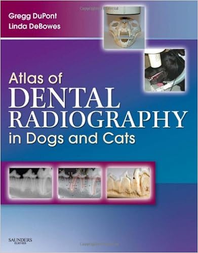
Is it ever applicable to diagnose and deal with oral and dental difficulties with out figuring out the whole quantity of the matter? With greater than 50% of anatomical buildings and linked pathologies situated lower than the gingivae and unseen to the attention, that is the fact with no using fine quality, effectively interpreted radiographs. Atlas of Dental Radiography in canine and Cats provides 1000s of tangible radiographic photographs, that are essentially categorised to facilitate actual id of ordinary and irregular gains. This helpful new atlas exhibits you precisely tips to correlate universal dental stipulations with radiographic symptoms. Radiographs also are in comparison aspect by means of part with real anatomical images to verify floor landmarks noticeable at the radiographs. right positioning options for generating diagnostic radiographs in addition to precious information and pitfalls whilst acquiring caliber radiographs are logically provided. This method is helping you produce continuously fine quality radiographs, sharpen your interpretive abilities, and with a bit of luck deal with quite a lot of dental problems.Presents the main logical and necessary method of dental and oral radiography, utilizing genuine anatomical images for actual medical correlationDepicts unique and color-labeled radiographs side-by-side for actual identity of standard and irregular structuresHelps either veterinarians and technicians take the very best radiographs, interpret them competently, make sound remedy judgements, and computer screen resultsProvides transparent, technical information for taking caliber radiographs and selecting artefacts and result of fallacious imaging process and picture developmentPresents transparent pictorial directions - from 2 angles - for proper positioning of the X-ray beam and intraoral filmsOffers new possibilities for accelerated specialist companies and sales on your practiceProvides facts of compliance with criteria of deal with clinical checklist documentation, assisting you legally guard yourself, your employees, and your perform
Read Online or Download Atlas of Dental Radiography in Dogs and Cats PDF
Best dentistry books
Книга Tyldesley's Oral medication Tyldesley's Oral MedicineКниги Медицина Автор: Anne box , Lesley Longman, William R. Tyldesley, Год издания: 2003 Формат: chm Издат. :Oxford college Press Страниц: 256 Размер: 20,7 ISBN: 0192631470 Язык: Английский0 (голосов: zero) Оценка:Firmly proven because the textbook of selection at the topic, Tyldesley's Oral medication is exclusive in its complete insurance at a degree compatible for either undergraduate and postgraduate dental scholars and practitioners.
The Moral Economy of AIDS in South Africa (Cambridge Africa Collections)
Fairly few humans have entry to antiretroviral remedy in South Africa. the govt justifies this on grounds of affordability, a view that Nicoli Nattrass argues is insulating AIDS coverage from social dialogue and the probabilities of financing a wide scale intervention. Nattrass addresses South Africa's contentious AIDS coverage from either an monetary and moral standpoint.
Sturdevant's Art and Science of Operative Dentistry
This finished textual content provides an in depth, seriously illustrated, step by step method of restorative and preventive dentistry. It attracts from either thought and perform, and is supported by means of vast scientific and laboratory learn. dependent upon the main that dental caries is a disorder, now not a lesion, the e-book offers either a radical figuring out of caries and an authoritative method of its therapy and prevention.
- Peterson's Principles of Oral and Maxillofacial Surgery
- Endodontics
- Practical Dental Local Anaesthesia (2nd Edition) (Quintessentials of Dental Practice, Volume 6; Oral Surgery & Oral Medicine, Volume 1)
- Teeth for Life for Older Adults (Quintessentials of Dental Practice, Volume 7; Prosthodontics, Volume 1)
- Health Promotion in Communities: Holistic and Wellness Approaches
Additional info for Atlas of Dental Radiography in Dogs and Cats
Example text
10. 11. 12. 13. indd 37 E F 4/25/08 3:22:06 PM 38 PART TWO RADIOGRAPHIC ANATOMY MAXILLARY MOLAR TEETH * 2 2 1 1 B A FIGURE 2-32 A, Normal radiolucency that can mimic a fracture of the maxillary tuberosity (arrows). B, Radiograph of a skull specimen using an angle and technique to reproduce the effect. This appearance is caused by summation effect (burn-out) below the radiodense zygomatic arch (asterisk). First molar (1), second molar (2). 2 * 1 A 1 2 * B FIGURE 2-33 Supernumerary molar teeth. A, Bassett hound with a maxillary third molar tooth (asterisk).
4. indd 35 Condensing osteitis indicating a chronic lesion. Loss of buccal plate from periodontitis Loss of both buccal and palatal bone in furcation Periapical and periradicular lucencies consistent with lesions of endodontic origin 4/25/08 3:22:05 PM 36 PART TWO RADIOGRAPHIC ANATOMY MAXILLARY MOLAR TEETH A B C FIGURE 2-31 Normal maxillary molar teeth. A, Radiograph of left maxillary molars. B, Buccal view of molar region of skull. C, Palatal view (mirror image) of molar region of skull. indd 36 4/25/08 3:22:05 PM CHAPTER 2 INTRAORAL RADIOGRAPHIC ANATOMY OF THE DOG 37 MAXILLARY MOLAR TEETH 5 7 12 7 3 12 5 7 1 13 6 8 4 3 6 1 12 11 2 10 9 D FIGURE 2-31, cont’d D, Same radiograph as A.
C, Two-year-old Toy Poodle. The dentin walls are thicker due to secondary dentin deposition. The apical sections of both roots are dilacerated with distinct distal angulation (arrows). This is the result of continued root development after complete eruption in a small-breed dog with a high tooth-to-bone ratio. D, Nine-year-old Dalmation mix. There is marked attenuation of the pulp chamber and root canal spaces. This molar tooth has a very distinct double-shadow due to a deep radicular groove on the distal aspect of the mesial root (arrows).


